Photography of the Unseen Part 1: Photomicrography
The first installment in our two-part series on extreme-scale photography
Products are chosen independently by our editors. Purchases made through our links may earn us a commission.
Photography is all about capturing images of the world in ways that challenge our natural perspective. As the photographer Alfred Stieglitz said, "In photography, there is a reality so subtle that it becomes more real than reality."
What's to say, then, about the realities that are impossible to see through the naked eye? Specifically, the microscopic world of cellular life and atomic matter? Can photography on the smallest of scales help us better understand life at the human level? Are these depictions of the infinitesimal somehow more "real" than the theories that describe them?
Those are some big questions, none of which we can answer, so let's just take a look at how we photograph really small things.
The Technology
When it comes to microscopy, it helps to have a sense of scale. Consider this: The smallest object visible to the human eye has a width of about 0.1 mm, or 100,000 nanometers (nm)—roughly the size of a paramecium. But it would still be very difficult to glimpse such an object, and anything smaller would require the use of a microscope.
Standard light microscopes are able to resolve anything larger than the wavelength of visible light, which is roughly 500 nm. That puts everything from cellular mitochondria and X chromosomes to red blood cells and baker's yeast well within the range of observation. However, atoms, proteins, and even viral cells are impossible to view through a standard light microscope—regardless of resolution.
Electron microscopes allow for observation of nano-scale objects by relying on the wavelengths of individual electron beams. While these machines can depict tiny viral cells and even atomic masses, they do so at a loss to resolution. Accordingly, researchers are working to develop a sort of "quantum microscope" capable of observing not just groups of atoms, but the precise orbital structure of individual particles.
For a truly mind-blowing representation of the scales involved, check out this interactive display from the Genetic Science Learning Center at the University of Utah.
The Science
Back in 2013, Aneta Stodolna, of the FOM Institute for Atomic and Molecular Physics (AMOLF) in the Netherlands, built a microscope that uses photoionization to directly map the electron orbit of an excited hydrogen atom. While low on resolution, the image is breathtaking—and it's the first direct observation of the orbital structure of a hydrogen atom.

In 2013, Dutch researchers used photoionization microscopy to map out the electron orbit of a hydrogen atom placed in an electrical field.
What makes this photograph so interesting is the fact that it offers a first-hand glimpse of a problem that's been plaguing physicists for decades: the quantum observer effect. Put simply, this theory holds that the observation of particles affects their very existence, rendering the position or momentum of electrons random and indecipherable.
However, Stodolna's microscope was able to depict the wave structure of a hydrogen atom's electron orbit while placed within a static electric field.
Researchers at Harvard University recently demonstrated a different method of photographing microscopic objects. These scientists probed the tiny magnetic fields produced by special kinds of bacteria that contain magnetic nanoparticles. This allowed them to produce images with a resolution display of just 400 nanometers.
{{amazon name="Practical Digital Photomicrography: Photography Through the Microscope for the Life Sciences", asin="1933952075", align="right"}} In theory, such technology could allow for real-time observation of internal cellular processes, such as cell death and division, as well as how cells are affected by disease. It could also lead to improved resolution and contrast in magnetic resonance imaging (MRI) technology.
Another fascinating breakthrough in photomicrography occurred in May of 2013, when researchers captured the first-ever high-resolution images of a molecule as it broke apart and reformed its chemical bonds. Scientists at the Lawrence Berkeley National Laboratory used an atomic force microscope to observe the phenomenon. Note the similarity of the molecule's actual appearance to that of the illustrations in your high school chemistry textbook.

With textbook clarity, this image taken by an atomic force microscope shows the position of individual atoms and bonds.
The Art
The value of photomicrography extends beyond mere science and technology. For decades, artists and photographers have relied on microscopic imaging to create beautiful displays of life at the microscopic level. Some manufacturers even have whole departments devoted to the field.
Nikon's Small World competition has been celebrating the beauty of light microscopy for 38 years. Past winners have included images of ink being injected into the vascular network of a chick embryo, and a water flea playing with a green alga.
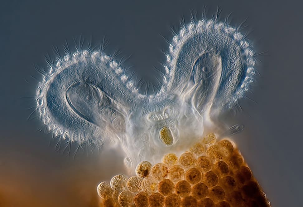
Olympus hosts a similar competition, called BioScapes, that deals specifically with life sciences. The below image is the competition's First Place winner from 2011. It depicts a tiny rotifer in the act of feeding. Interestingly, these creatures, also known as "wheel animalcules," were first described in 1702 by Antonie van Leeuwenhoek—the man often referred to as the "first microbiologist," thanks to his tremendous improvements to the light microscope.
The microscopic world is, in a sense, its own kind of universe, replete with strange organisms and physical principles that make the head spin. However, it is perhaps just as illuminating to turn your head up as it is down. With that in mind, check out part 2 in our series on photography of the unseen: astrophotography.
Related Video
{{brightcove '4170904179001'}}

